キーワード: 腫瘍特異的ヘム- ポルフィリン代謝、光化学診断法、がん治療法、活性酸素
日本をはじめとする先進国では高齢化を迎えて、脳血管障害や心臓冠状動脈疾患等の原因である動脈硬化の患者が急速に増加しています。動脈硬化の治療には血小板を固めないようにアスピリン等の薬物を投与します。その為、この患者ががんなどの別の疾患を発症すると、出血を伴う手術が困難となります。このような背景の下、現在見直されているがん治療としてレーザーなどを用いた光化学療法があります。低侵襲で出血の少ない治療が求められているのです。
がんの光化学療法では、がん細胞に集まるポルフィリンという蛍光色素にレーザーを当てて活性酸素を発生させ腫瘍細胞を壊します。正常細胞ではポルフィリンは鉄分子と結合しヘムという物質に代謝されますが、がん細胞では代謝酵素が失活しているためにポルフィリンが蓄積します。本ユニットではこの蛍光物質の蓄積機序を解明することで、がんの診断・治療に役立てようとしています。ポルフィリン前駆体の増減により蛍光物質の蓄積を制御し、蛍光を強めてがんの領域診断に役立てることや、蛍光物質にレーザーを照射した際に発生する多量の活性酸素を利用してがん細胞を特異的に殺傷することが実現します。
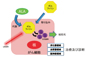
図1:ALA はポルフィリンとしてがん細胞に蓄積する。これにレーザーを当てると細胞傷害性の高い活性酸素が発生する。
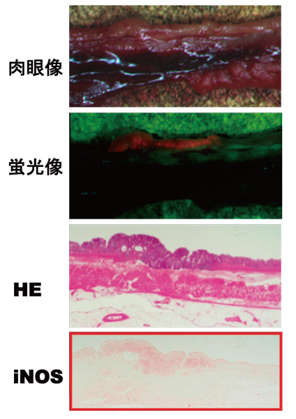
図2:がん手術検体の組織像(赤枠は組織免疫染色像)蛍光像で赤色蛍光を放っている部分と活性酸素種を産生するiNOS の局在が一致している。赤色蛍光部ががん部位である事はHE 染色から確認する事が出来る。
様々な領域において、がん代謝に着目した光化学診断が始まっている
本ユニットは脳神経外科、消化器内科、腎泌尿器外科に横断したチームです。脳神経外科領域では、脳腫瘍の手術の際に光化学診断を用います。患者がアミノレブリン酸(ALA)というポルフィリンの前駆体を飲むことで腫瘍患部が光り、それを手掛かりに顕微鏡を見ながら光った部位だけを切除します。
泌尿器領域では、ALA の摂取後の尿を調べます。尿中にALA から生成されたポルフィリンが入っていたら、尿が赤く光ることで膀胱がんのスクリーニングが出来ます。また赤色を頼りに膀胱鏡で治療を試みます。
がんの代謝を見る診断・診療は世界の中でも日本がリードしている領域であり、更に発展させていくことを目指しています。
光化学診断の可能性を広げる新しい取組み
新しいチャレンジとして循環腫瘍細胞の分離に取組んでいます。循環腫瘍細胞は血管の中を循環しており、がんの転移メカニズムや薬剤耐性機序などを紐解くものと言われていますが、この分離・培養が難しく世界中の研究者が挑んでいます。本ユニットでは、細胞表面に標識を付けて分離するのではなく、ALA を作用させ光った細胞を回収するという方法の開発にベンチャー企業と共に取組んでいます。また、異分野とのコラボレーションにより、新しい光線力学療法の材料を藻等の植物から合成するといったことにも取組んでいます。
社会への貢献・実績
- 日本酸化ストレス学会フリーラジカルサマースクール開催(2012 ~ 2015 年度毎年開催)
取材:平成27年4月27日
Detection and treatment of cancer using cancer cell metabolism
Unit members : 及川 剛宏 Kaneko Tsuyoshi Nishiyama, Hiroyuki Nagasaki, Yukio
Unit name: Clinical Research Unit for Tumor Specific Heme Metabolisms
Key words: tumor-specific heme-porphyrin metabolism, photochemical diagnosis method, cancer treatment method, reactive oxygen species
Due to the population aging in developed countries, such as Japan, patients with atherosclerosis, which causes cerebrovascular or coronary heart diseases, have been rapidly increasing. As treatment for atherosclerosis, drugs, such as aspirin, are administered to prevent platelets from forming clots. Therefore, when such patients develop other diseases, such as cancer, surgery accompanied by bleeding is difficult. With such a background, photochemical therapy using light sources, such as lasers, which is minimally invasive and causes minimal bleeding, has been attracting attention.
In photochemical therapy for cancer, laser light is applied to porphyrin as a fluorescent dye accumulating in cancer cells to generate reactive oxygen species for tumor cell destruction. In normal cells, porphyrin binds to iron molecules and is metabolized into heme. In cancer cells, due to the inactivation of the enzyme for its metabolism, porphyrin accumulates. Our unit aims to clarify the mechanism of the accumulation of this fluorescent material for cancer diagnosis and treatment. The control of the accumulation of the fluorescent material by the increase or decrease of the porphyrin precursor and enhancement of the fluorescence can contribute to cancer diagnosis. In addition, cancer cells can be specifically killed using a large amount of reactive oxygen species generated at the time of laser irradiation on the fluorescent material.
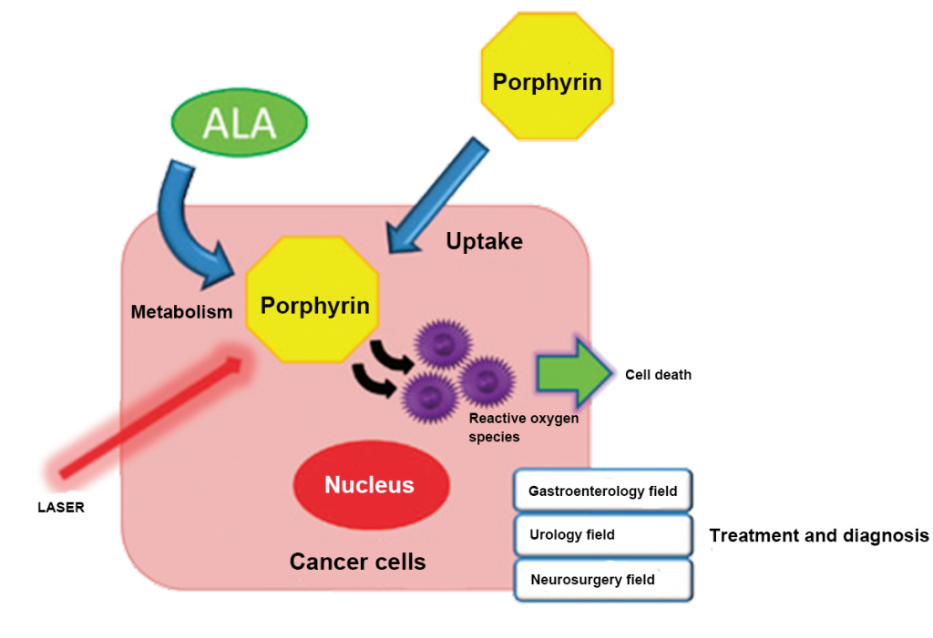
Fig. 1: ALA accumulates as porphyrin in cancer cells. Laser irradiation on porphyrin results in the generation of highly cytotoxic reactive oxygen species.
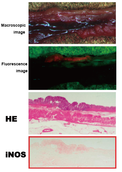
Fig. 2: Histological images of a surgical specimen of cancer (red frame: immunohistological staining)
Photochemical diagnosis focusing on cancer metabolism is beginning in various fields
This unit is a cross-functional team consisting of the Departments of Neurosurgery, Gastroenterology, and Urology. In the Department of Neurosurgery, photochemical diagnosis is used for brain tumor surgery. Patients drink a solution containing aminolevulic acid (ALA) as a porphyrin precursor. The tumor glows, and only the glowing area is resected under a microscope.
In the urology field, urine after oral ALA administration is examined. If porphyrin formed from ALA is present in the urine, urine glows red, which allows bladder cancer screening. In addition, red color-guided treatment is performed using a cystoscope.
Japan leads the world in the diagnosis and treatment using cancer metabolism. We aim to achieve further development.
New approaches to expand the possibilities of photochemical diagnosis
As a new challenge, we are attempting to separate circulating tumor cells. Circulating tumor cells circulate in the bloodstream, and are considered to be the key for the elucidation of the mechanisms of tumor metastasis and drug tolerance. The separation and culture of these cells are difficult, and researchers throughout the world are making efforts. In cooperation with a venture company, our unit is developing a method, in which ALA is allowed to react with cells, and glowing cells are collected, unlike methods in which a marker is attached to the cell surface, and cells are separated. In addition, we are developing new materials for photodynamic therapy using plants such as algae in collaboration with other fields.
Social contributions and achievements
- Free Radical Summer School of the Society for Free Radical Research Japan was held (annually in the fiscal years 2012-2015).
Interviewed on April 27, 2015